
FUNDRAISING FOR A CURE
Donating is simple, fast and totally secure. Your details are safe with us and we will never sell them
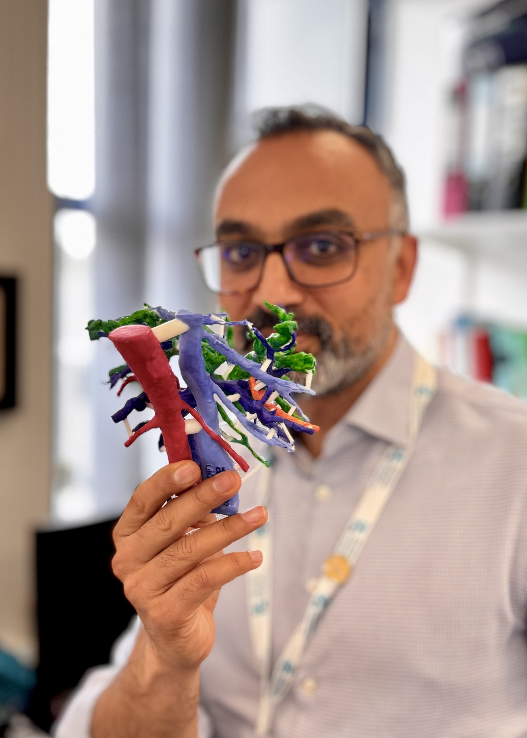
Surgeons in Southampton are the first in the UK to begin using 3D printed models of patients’ livers to help them perform a complex cancer operation.
Converting the data available from CT and MRI scans of patients with hilar cholangiocarcinoma – a type of bile duct cancer – into 3D models will enable better planning and decision-making prior to surgery.
As the cancer occurs in the bile ducts that lead out of the liver and join with the gallbladder, it is frequently difficult for surgeons to establish whether a tumour can be safely removed or not until the operation is underway.
As a result, this can lead to “irreversible steps” being taken that may result in poor short and long-term outcomes for patients.
By using 3D printed models tailored to each patient, clinicians will be able to assess the tumour and its close attachments such as blood vessels and bile ducts at scale pre-surgery.
The project will be led by Mr Arjun Takhar, a consultant hepatobiliary and pancreatic cancer surgeon at University Hospital Southampton, who is part of a team of experts supported by PLANETS Cancer Charity, which is funding the pilot study and has received a £2,000 grant from The Hospital Saturday Fund to put towards the initiative.
PLANETS helps patients with pancreatic, liver, colorectal, abdominal (oesophageal and gastric) and neuroendocrine cancer by funding patient support groups, innovative treatments and research.
The charity primarily serves the needs of the regional population living across Hampshire, Wiltshire, Dorset, the Isle of Wight, the Channel Islands and West Sussex, it is increasingly involved in national and international clinical and research initiatives.
“3D printed models are increasingly being used to help with decision-making before and during surgery and to better understand the anatomical relationships of tumours within organ structures,” explained Mr Takhar.
“With this particularly challenging liver/bile duct cancer – hilar cholangiocarcinoma – sometimes we are unable to tell until the very last minute whether the tumour can be safely removed or not.
“Sometimes surgeons will have taken an irreversible step and then found out that the tumour cannot be removed completely which results in poor outcomes for patients in the short and long-term.”
He added: “3D printing offers the advantage of assessing the tumour and its close attachments such as blood vessels and bile ducts in a scale model prior to performing the operation itself.
“The aim of the pilot project is to assess whether this allows for assessment of operability to be made without opening the patient up and also whether assessment with a model is better than simply looking the patient’s CT and MRI scans.
“There is also an added advantage that a 3D model would also help in teaching trainee surgeons the nuances of liver anatomy in relation to these complex tumours.
“This is a unique opportunity to use a novel technology to help patients with a difficult disease and we foresee adoption of the technology in patients with other liver tumours too.”
READ ABOUT HOW FUNDS ARE USED
-
PLANETS Funds New Wellbeing Room for Liver Ward at UHS
PLANETS funds new wellbeing room for liver patients at University Hospital Southampton A new wellbeing room funded by PLANETS Cancer Charity has been opened for patients with liver diseases at University Hospital Southampton. The facility, located on the gastroenterology and hepatology ward, provides a quiet space for patients and a place for private conversations which […]
-
PLANETS funds novel 3D printed livers to aid complex surgery
Surgeons in Southampton are the first in the UK to begin using 3D printed models of patients’ livers to help them perform a complex cancer operation. Converting the data available from CT and MRI scans of patients with hilar cholangiocarcinoma – a type of bile duct cancer – into 3D models will enable better planning […]
-
PLANETS Charity Welcome Coach Alison Bailey to their Support Team
Here at PLANETS we understand how daunting a cancer diagnosis can be. We know firsthand the transformative power of support throughout the cancer journey and have experienced ourselves the highs and lows, the triumphs and setbacks and the profound impact they can have on your life. We are very happy to be welcoming Alison Bailey, […]
-
PLANETS completes £1 million fundraising campaign to purchase pioneering radiotherapy machine
PLANETS has thanked its supporters after completing a £1 million fundraising campaign and making the final payment for a pioneering radiotherapy machine. PLANETS Cancer Charity has been fundraising for the past six years to fully purchase Mobetron, a revolutionary mobile device which delivers radiotherapy during surgery – known as intraoperative radiotherapy (IORT). When the charity […]
-
PLANETS Receive £10,000 Grant from The Hospital Saturday Fund
Thank you to John Greenwood, Trustee of the The Hospital Saturday Fund, for visiting us on Monday and bringing this incredible grant of £10,000 to support our IORT (Intra Operative Radiotherapy) machine. This will enable us to continue delivering this lifesaving treatment at University Hospital Southampton – the only centre in the UK currently offering […]
-
PLANETS Fund ‘New View’ Psychotherapy Course for NET Patients
PLANETS have funded a psychotherapy programme, after a successful pilot course, to help NET patients see their diagnosis in a ‘new view’. We all know that different people deal with their neuroendocrine cancer diagnosis very differently. Initially there are lots of scary tests and Doctor appointments. Then there are lots of talk about […]
-
Head and Neck Cancer Patient Treated with IORT in UK First
A woman from Salisbury has become the first patient in the UK with head and neck cancer to receive radiotherapy in the operating theatre during surgery to remove a recurrent tumour. Jayne Garrett, 53, underwent major surgery at University Hospital Southampton on 25 April after suffering a recurrence of her cancer following conventional treatment […]
-
PLANETS Host the 1st National IORT Symposium
It was an honour to present the fantastic work and great initial results to medics from all over the country at our 1st National IORT (intra-operative radiotherapy) Symposium on Friday June 21st at University Hospital Southampton. After a lot of hard work and fundraising in order to bring IORT to the UK for the […]
-
PLANETS Cancer Charity and the Robert White Trust, Fund a new regional NET Cancer Dietitian
A dietitian funded by PLANETS Cancer Charity and the Robert White Trust Fund is due to start work to develop and provide a comprehensive dietetic service to patients diagnosed with Neuroendocrine Cancer (NETs). Ruth Lee will be based at the Dorset NET service, which is part of the Wessex European Neuroendocrine tumour society centre […]
-
Southampton Secure £200k from The Robert White Trust for NETs Research
PLANETS are excited to share the news that University Hospital Southampton have secured £200,000 funding from the Robert White Trust for a research project that aims to gain a better understanding of NET biology which may lead to a greater range of treatment options for patients with this rare disease. Robert White was a former […]
-
Bowel cancer patient becomes first in UK to be treated with IORT
We are thrilled and proud to update our supporters that a Southampton bowel cancer patient has become the first in the UK to receive radiotherapy during surgery using the IORT machine that PLANETS has funded. he 58-year-old male, who completed a combination of conventional chemotherapy and radiotherapy in August, underwent major surgery at Southampton […]
-
IORT Launch is a Success!
SOUTHAMPTON CLINICIANS PIONEER USE OF REVOLUTIONARY CANCER DEVICE PLANETS founders Neil Pearce and Brian Stedman, together with fund manager Layla Stephen successfully launched our long awaited IORT machine last night. The Mobetron is the first portable system able to administer the treatment in this way – known as intraoperative radiotherapy (IORT) – and will […]
-
IORT Arrives at Southampton!
PLANETS are thrilled to announce that the eagle (finally) has landed! 2 tons of intra-operative radio-therapy machine (IORT) arrived from the USA this week – currently undergoing acceptance testing. This is the UK’s first ever mobile electron beam radiotherapy machine for treating cancers during surgery. Thanks to everyone who has supported our fundraising which has […]
-
Southampton Aiding Research into Earlier Diagnosis for Pancreatic Cancer
You may have notice that Southampton was mentioned in recent media stories regarding The National Tumour Bank that is carrying out important pancreatic cancer research? Colin Johnson, Professor of Surgical Sciences at University of Southampton, has this to say about the project: ‘The National Tissue Bank is a major development in pancreatic cancer research. Several […]
-
Sainsbury’s Hedge End Donate Digital Radios
A big thank you to Sainsbury’s Hedge End for donating two digital radios to PLANETS to be used in the lead lined Gamma Scanner Rooms at Southampton General. Pictured presenting the radios to staff in the scanner rooms are PLANETS Fund Manager, Layla Stephen, and Dr Brian Stedman.
-
PLANETS to Fund Pancreatic Cancer Research at Southampton
PLANETS Charitable Fund are excited to have committed to provide a grant over a two year period for a pancreatic research project: ́The role of Eps8 in αvβ6-dependent functions in pancreatic cancer invasion’ to be undertaken by Dr Jo Tod. Pancreatic adenocarcinoma (PC) has one of the worst outcomes of any cancer; only 3.7% of patients […]
-
First Patient Receives PRRT at Southampton thanks to PLANETS
[social_button button=”facebook” flayout=”standard ” fwidth=”450″ faction=”like” fcolorsheme=”light”] UHS magazine Connect featured a fantastic article about NET patient Maureen McKenzie who, in July 2013, became the first Southampton patient to receive PRRT (peptide receptor radionuclide therapy). The equipment for this treatment was jointly funded by UHS and PLANETS showing just how important and valuable the […]
-
Improving Patient Services Across the Region
[social_button button=”facebook” flayout=”standard ” fwidth=”450″ faction=”like” fcolorsheme=”light”] Pancreatic cancer affects over 8000 patients each year in the uk and remains the fifth biggest cancer killer in the UK, despite this its has been underfunded for many years and currently receives just 1% of total cancer research funding in the UK. NETs are a rare form […]

Subscribe to our Newsletter
Get informed about the latest news straight to your inbox